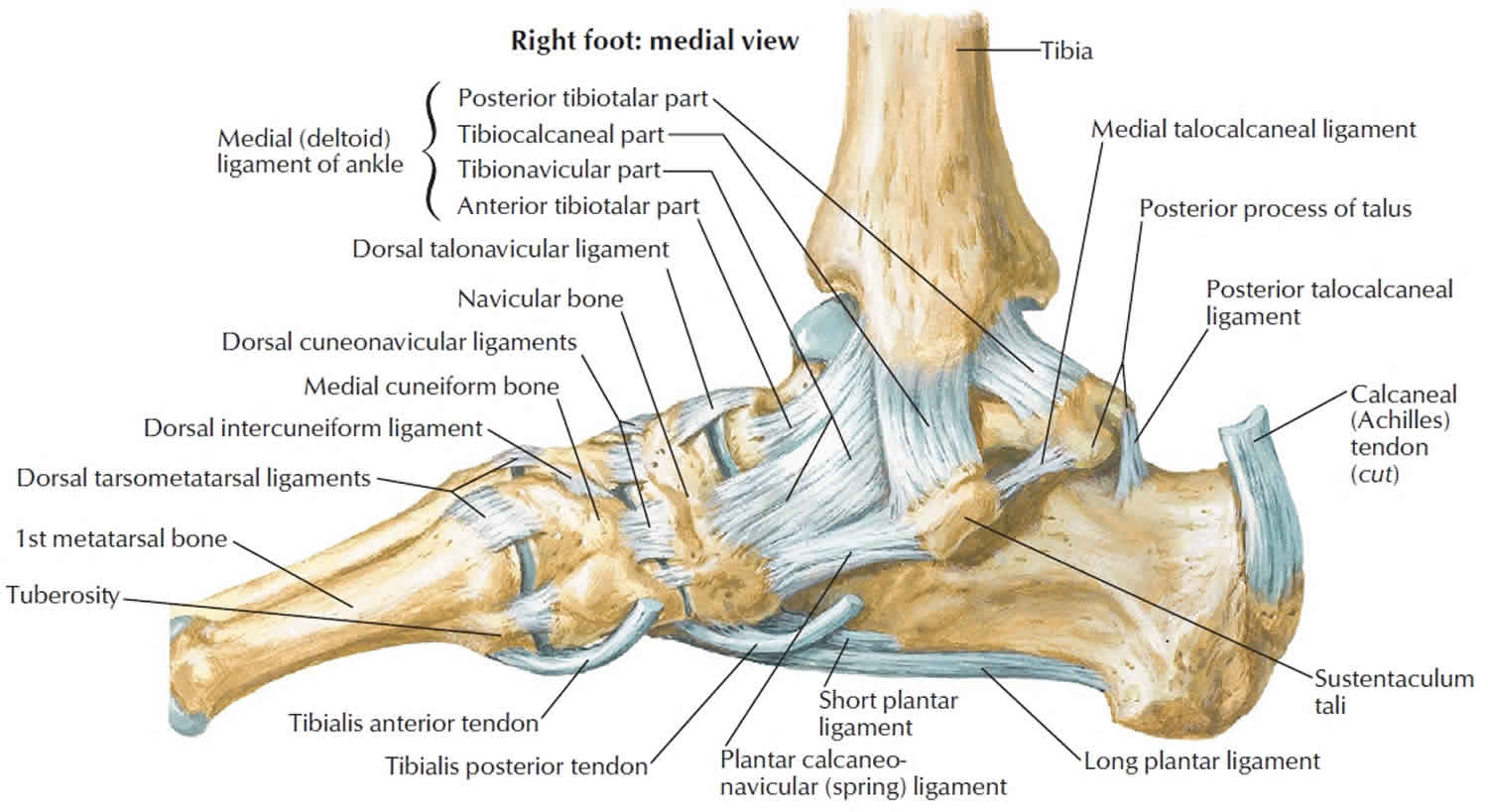
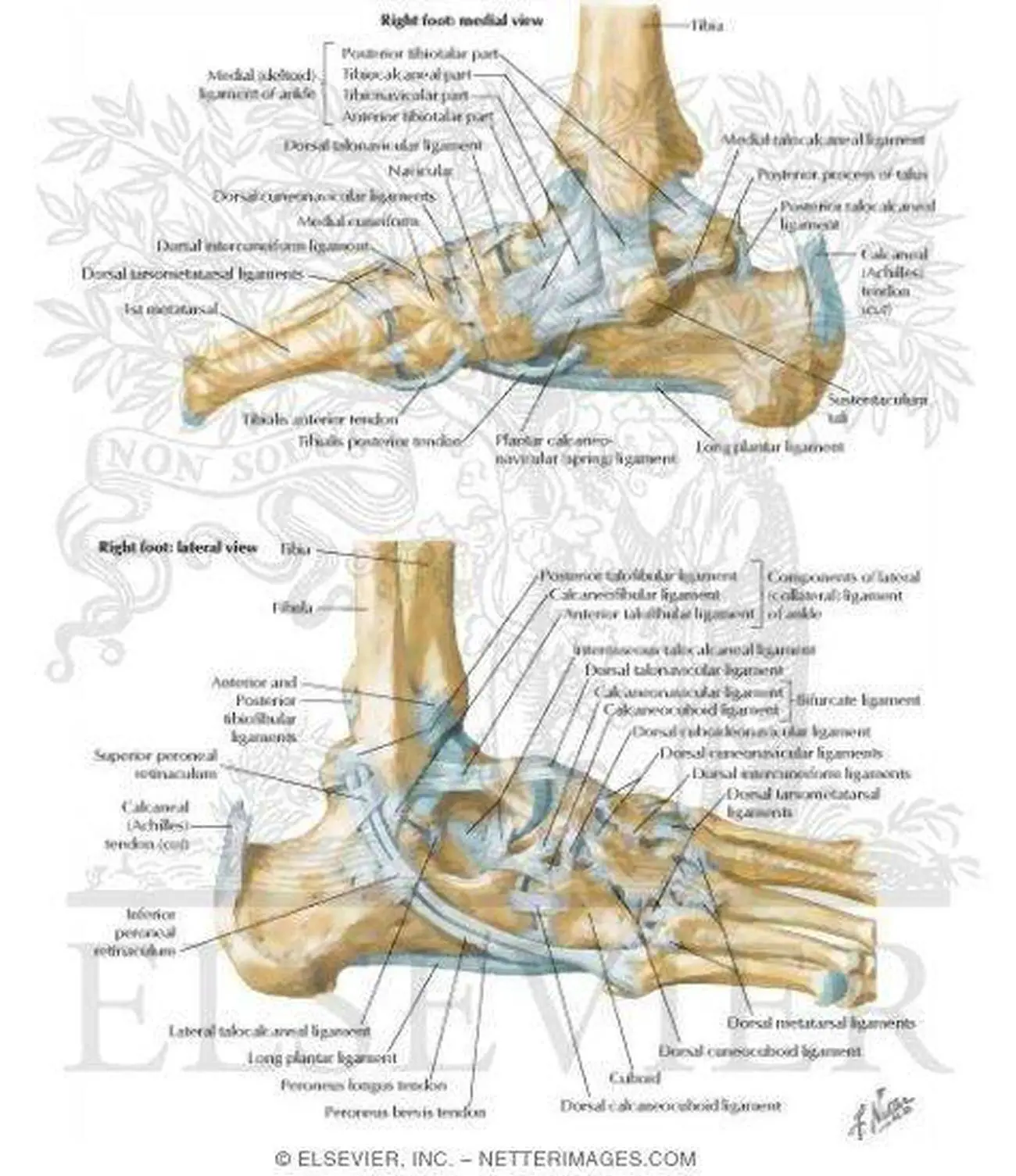
Tendon Diagram Foot / Pictures Of Ankle Joint Ligaments - This is meant for educational purposes only.. If you would like to learn all the parts of the foot structure, you have come to the right place. The thigh and leg bones articulate at the knee joint that is protected and enhanced by the patella bone that supports the quadriceps tendon. There are a whole range of structures e.g. Bones, muscles, tendons and nerves which will each give slightly different foot pain symptoms. The bones of the foot include the tarsus, metatarsus, and phalanges.
Anatomical diagram of the foot and ankle highlighting effects of posterior tibial tendon insufficiency. Here you can see the tendons that extend down the top of your foot toward your toes, allowing you to curl your toes upward if need be. A foot pain diagram is a great tool to help you work out what is causing your ankle and foot pain. If you would like to learn all the parts of the foot structure, you have come to the right place. Many tears do not completely sever the tendon.
Calcaneus bone anatomy, function, calcaneus pain ... from healthjade.com Posterior tibial tendon dysfunction (pttd) is a condition caused by changes in the tendon, impairing its ability to support the arch. A complete tear will split the tendon into two pieces. Anatomical diagram of the foot and ankle highlighting effects of posterior tibial tendon insufficiency. The bones of the foot include the tarsus, metatarsus, and phalanges. Jul 05, 2018 · the foot diagram has a complex structure made up of bones, ligaments, muscles, and tendons. The posterior tibial tendon serves as one of the major supporting structures of the foot, helping it to function while walking. As the damage progresses, the tendon can completely tear, sometimes when lifting a heavy object. The neck of the femur terminates at the lesser and greater trochanter prominences.
Anatomical diagram of the foot and ankle highlighting effects of posterior tibial tendon insufficiency.
Understanding the structure of the foot is best done by looking at a foot diagram where the anatomy has been labeled. Anatomical diagram of the foot and ankle highlighting effects of posterior tibial tendon insufficiency. Jun 07, 2019 · this results in collapse of the arch of the foot (commonly called flatfoot or flat foot), along with foot and sometimes ankle deformities that can become debilitating or disabling in later stages. Top (dorsal) view of foot & ankle number 1 and 2: This results in flattening of the foot. Placing heel lifts inside the shoes or wearing shoes with a moderate heel takes stress off the achilles tendon when walking and may reduce symptoms. A foot pain diagram is a great tool to help you work out what is causing your ankle and foot pain. The foot may be placed in a splint at night to keep it in a position that helps reduce tightness of the calf muscle. There are a whole range of structures e.g. Jul 05, 2018 · the foot diagram has a complex structure made up of bones, ligaments, muscles, and tendons. This is meant for educational purposes only. Bones, muscles, tendons and nerves which will each give slightly different foot pain symptoms. Match the corresponding numbers on the foot diagram below for a list of conditions that may be causing your foot and ankle pain.
Many tears do not completely sever the tendon. This results in flattening of the foot. Top (dorsal) view of foot & ankle number 1 and 2: The bones of the foot include the tarsus, metatarsus, and phalanges. Match the corresponding numbers on the foot diagram below for a list of conditions that may be causing your foot and ankle pain.
Muscles of the Lower Extremities. Muscular system from encyclopedia.lubopitko-bg.com A complete tear will split the tendon into two pieces. Many tears do not completely sever the tendon. Bones, muscles, tendons and nerves which will each give slightly different foot pain symptoms. Placing heel lifts inside the shoes or wearing shoes with a moderate heel takes stress off the achilles tendon when walking and may reduce symptoms. There are a whole range of structures e.g. The thigh and leg bones articulate at the knee joint that is protected and enhanced by the patella bone that supports the quadriceps tendon. The abductor muscles attach to the greater trochanter while the lesser trochanter is the site of attachment for the iliopsoas tendon. The foot may be placed in a splint at night to keep it in a position that helps reduce tightness of the calf muscle.
Jan 01, 2019 · the hip joint is attached to the posterior surface of the femoral neck via the capsular ligament.
Bones, muscles, tendons and nerves which will each give slightly different foot pain symptoms. A foot pain diagram is a great tool to help you work out what is causing your ankle and foot pain. Chloe wilson bsc(hons) physiotherapy reviewed by: The foot may be placed in a splint at night to keep it in a position that helps reduce tightness of the calf muscle. There are a whole range of structures e.g. Jul 05, 2018 · the foot diagram has a complex structure made up of bones, ligaments, muscles, and tendons. Here you can see the tendons that extend down the top of your foot toward your toes, allowing you to curl your toes upward if need be. Jan 01, 2019 · the hip joint is attached to the posterior surface of the femoral neck via the capsular ligament. Jun 07, 2019 · this results in collapse of the arch of the foot (commonly called flatfoot or flat foot), along with foot and sometimes ankle deformities that can become debilitating or disabling in later stages. In many cases, torn tendons begin by fraying. Placing heel lifts inside the shoes or wearing shoes with a moderate heel takes stress off the achilles tendon when walking and may reduce symptoms. The posterior tibial tendon serves as one of the major supporting structures of the foot, helping it to function while walking. The abductor muscles attach to the greater trochanter while the lesser trochanter is the site of attachment for the iliopsoas tendon.
There are a whole range of structures e.g. Chloe wilson bsc(hons) physiotherapy reviewed by: If you would like to learn all the parts of the foot structure, you have come to the right place. Biceps tendon tears can be either partial or complete. Jun 07, 2019 · this results in collapse of the arch of the foot (commonly called flatfoot or flat foot), along with foot and sometimes ankle deformities that can become debilitating or disabling in later stages.
Pictures Of Ankle Joint Ligaments from healthiack.com The posterior tibial tendon serves as one of the major supporting structures of the foot, helping it to function while walking. Many tears do not completely sever the tendon. Here you can see the tendons that extend down the top of your foot toward your toes, allowing you to curl your toes upward if need be. Top (dorsal) view of foot & ankle number 1 and 2: The foot may be placed in a splint at night to keep it in a position that helps reduce tightness of the calf muscle. A complete tear will split the tendon into two pieces. A foot pain diagram is a great tool to help you work out what is causing your ankle and foot pain. The bones of the foot include the tarsus, metatarsus, and phalanges.
Jan 01, 2019 · the hip joint is attached to the posterior surface of the femoral neck via the capsular ligament.
The abductor muscles attach to the greater trochanter while the lesser trochanter is the site of attachment for the iliopsoas tendon. If you would like to learn all the parts of the foot structure, you have come to the right place. As the damage progresses, the tendon can completely tear, sometimes when lifting a heavy object. Here you can see the tendons that extend down the top of your foot toward your toes, allowing you to curl your toes upward if need be. There are a whole range of structures e.g. Bones, muscles, tendons and nerves which will each give slightly different foot pain symptoms. A complete tear will split the tendon into two pieces. The foot may be placed in a splint at night to keep it in a position that helps reduce tightness of the calf muscle. Jun 07, 2019 · this results in collapse of the arch of the foot (commonly called flatfoot or flat foot), along with foot and sometimes ankle deformities that can become debilitating or disabling in later stages. Biceps tendon tears can be either partial or complete. Placing heel lifts inside the shoes or wearing shoes with a moderate heel takes stress off the achilles tendon when walking and may reduce symptoms. This is meant for educational purposes only. Posterior tibial tendon dysfunction (pttd) is a condition caused by changes in the tendon, impairing its ability to support the arch.
A foot pain diagram is a great tool to help you work out what is causing your ankle and foot pain tendon diagram. Understanding the structure of the foot is best done by looking at a foot diagram where the anatomy has been labeled.


0 Komentar